Cephalometric imaging is one of the most important techniques in maxillofacial radiography, providing precise information about skeletal and dental structures. In this article, you will learn about the details of this method, its applications in orthodontics and jaw surgeries, and the process of its implementation.
If you need more information for treating jaw and facial abnormalities or planning orthodontic treatments, this article is for you. We also address common questions you might have, such as: What are the applications of cephalometry? Who is this method suitable for? And how does it differ from other techniques? This knowledge is essential to help you make the best treatment decisions.
Questions Answered in This Article:
- Who needs cephalometric imaging?
- Does cephalometric imaging have any risks?
- How long does it take to perform cephalometric imaging?
- What is the difference between cephalometric imaging and panoramic imaging?
- Why do doctors recommend cephalometric imaging for orthodontics?
For detailed answers to these questions and to gain precise information, we recommend reading this article thoroughly.
Cephalometric imaging is a specialized imaging technique in dentistry and orthodontics used to analyze craniofacial structures, including bones and soft tissues of the face. First introduced in 1931 by Broadbent and Hofrath, it became a key tool for the accurate diagnosis of orthodontic issues. Cephalometry allows for the examination of the relationships between skeletal and dental structures using two-dimensional and three-dimensional radiographic images. Its applications include diagnosing malocclusions, planning orthodontic treatments, and assessing craniofacial growth.
In recent years, advanced technologies such as cone-beam computed tomography (CBCT) and three-dimensional analysis have expanded the applications of cephalometry. These techniques offer greater precision in locating anatomical landmarks and analyzing long-term changes. Additionally, digital tools and advanced software are being utilized to enhance the efficiency of cephalometric analysis, enabling faster and more accurate identification of key points. These advancements have not only improved diagnostic and treatment planning processes but also facilitated clinical research in craniofacial growth and development.
What is cephalometric imaging, and why is it important?
Cephalometric imaging is a specialized technique in maxillofacial radiography that provides accurate images of the skeletal and soft structures of the jaw, face, and skull. This method is performed using X-rays and is primarily used in planning orthodontic treatments, jaw and facial surgeries, and evaluating skeletal abnormalities. Cephalometric imaging allows doctors to meticulously examine structural changes and design appropriate treatment plans.
The significance of this method lies in its ability to provide two-dimensional images that not only include a lateral view of the face but also reveal internal structures such as sinuses, jaw joints, and the relationship between teeth and jawbones. For this reason, the applications of cephalometry are extensive, and specialists rely on it to diagnose growth abnormalities and plan complex surgeries.
This technique is also recognized as a standard tool in orthodontics and reconstructive surgeries, offering high precision in measuring and analyzing skeletal and dental proportions. In many cases, cephalometric imaging serves as the first step in diagnosing facial and jaw abnormalities, helping physicians deliver precise, data-driven treatments.
Key Applications of Cephalometric Imaging in Jaw and Facial Treatments
The applications of cephalometry in jaw and facial treatments are extensive, making it a cornerstone diagnostic tool due to its ability to provide precise information about cranial structures. In orthodontics, cephalometric imaging is used to evaluate the position of teeth and their relationship with the upper and lower jaws. This data helps orthodontists adjust braces to achieve the best possible outcomes.
In jaw and facial surgeries, this technique serves as an essential tool for accurate planning. Surgeons can analyze structural abnormalities of the jaw and face and predict necessary adjustments. Moreover, maxillofacial radiography using cephalometric techniques aids in assessing the effects of previous treatments and correcting the alignment of teeth.
Another significant application of cephalometric imaging is in addressing speech disorders and breathing problems. This method provides valuable insights into the relationship between the tongue, larynx, and jaw structure, playing a critical role in diagnosing and treating these conditions.
How is Cephalometric Imaging Performed?
The process of cephalometric imaging is straightforward and painless but requires high precision. During the procedure, the patient must keep their head in a specific position to allow the equipment to capture a clear and accurate image of the cranial and jaw structures. The images are taken in two dimensions, typically including a lateral view of the head and face.
Before imaging, the doctor may ask the patient to remove all metal objects such as glasses or jewelry to ensure the image quality is not compromised. The X-ray machine is positioned at a specific angle, and the patient must remain still for a few seconds. At this stage, the applications of cephalometry become evident, as the images allow doctors to analyze the precise structure of the jaw and skull.
A key feature of maxillofacial radiography using cephalometric techniques is its accuracy in showing the relationship between teeth, the jaw, and soft tissues. The process takes only a few minutes and typically involves minimal radiation, making it safe for repeated use throughout treatment.

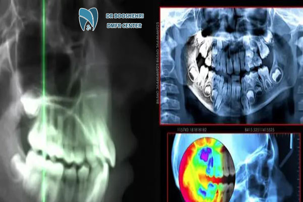
Cephalometric imaging is a vital technique in maxillofacial radiography. Discover the applications of cephalometry in diagnosing abnormalities
Comparison of Cephalometric Imaging with Other Maxillofacial Radiography Methods
Cephalometric imaging, compared to other maxillofacial radiography techniques like panoramic imaging or CBCT, has its unique advantages and limitations. While panoramic images are useful for providing a comprehensive view of dental and jaw structures, cephalometry offers more detailed information about skeletal proportions and the relationship of teeth to the skull.
CBCT, known for its 3D imaging and high resolution, is typically used for diagnosing more complex issues. However, it comes with higher costs and radiation exposure. In contrast, the applications of cephalometry are primarily focused on evaluating dental and skeletal abnormalities. Due to its lower cost and faster execution, it is often a more practical option for many patients.
Nonetheless, each method has its value, and depending on the patient’s needs and the type of treatment required, doctors may combine these techniques to achieve the best outcomes.
Who Needs Cephalometric Imaging?
Cephalometric imaging is beneficial for various patient groups. It is particularly recommended for individuals planning orthodontic treatments or jaw and facial surgeries. Teenagers undergoing growth changes are among the primary beneficiaries of this technique.
Additionally, patients with severe jaw, dental, or speech abnormalities can greatly benefit from maxillofacial radiography using cephalometry. Even individuals requiring evaluation of jaw and facial structures due to breathing problems or sleep disorders can take advantage of the applications of cephalometry in diagnosing such issues.
The Impact of Cephalometric Imaging on Diagnostic and Treatment Accuracy
One of the key reasons cephalometric imaging has gained attention in medical and dental fields is its significant contribution to improving diagnostic and treatment accuracy. This technique provides comprehensive and precise information, enabling healthcare professionals to make the best treatment decisions.
The images produced by cephalometric imaging allow doctors to anticipate necessary changes in dental and jaw structures and simulate potential treatment outcomes. In orthodontics, the applications of cephalometry include precise brace adjustments and treatment progress evaluations. In jaw and facial surgeries, this method serves as a tool for detailed analysis and surgical outcome prediction.
Ultimately, the use of maxillofacial radiography with cephalometric techniques dramatically reduces treatment errors, ensuring better results for patients.
Answers to Common Questions About Cephalometric Imaging
Who Needs Cephalometric Imaging?
Cephalometric imaging is essential for individuals undergoing orthodontic treatments, those with jaw and facial abnormalities, or patients preparing for jaw surgeries. This method helps doctors analyze jaw structures, teeth, and their relationships to develop precise treatment plans.
Is Cephalometric Imaging Harmful?
Cephalometric imaging uses X-rays, but the radiation dose is very low and considered safe. This procedure is entirely safe for patients, even with repeated use. However, if you are pregnant or have specific conditions, inform your doctor beforehand.
How Long Does Cephalometric Imaging Take?
The process is very quick, usually taking less than five minutes. The patient remains in a fixed position while a two-dimensional image is captured, either of the side or front view of the face. Results are also prepared promptly.
What is the Difference Between Cephalometric Imaging and Panoramic Imaging?
Panoramic imaging provides a comprehensive view of all teeth, jaws, and surrounding bones, while cephalometric imaging focuses on cranial structures and the relationship between the jaws and teeth to the skull. Cephalometry is mainly used for orthodontic planning and specialized jaw and facial surgeries.
Why Do Doctors Recommend Cephalometric Imaging for Orthodontics?
Cephalometric imaging provides detailed information about the position of teeth, upper and lower jaws, and their relationship to the skull. This data helps orthodontists plan the precise movement of teeth and improve jaw alignment, ensuring more accurate and effective treatment.
Why Should You Rely on Dr. Boushehri’s Expertise?
For accurate diagnoses and professional planning of orthodontic treatments or jaw and facial surgeries, visiting a specialized radiology center is crucial. Dr. Boushehri, a renowned specialist in oral, jaw, and facial radiology, brings extensive experience and utilizes the latest technologies to deliver the best diagnostic services.
At Dr. Boushehri’s specialized imaging center, you can benefit from high-quality, professional services and book a consultation if needed. Additionally, if you have any questions, you can ask them in the comments section and receive expert answers. Choosing Dr. Boushehri ensures precision and quality in diagnosis.


